Achieving the Highest Resolution Protein Structure Through Advanced X-Ray Diffraction Techniques
Researchers have determined the structure of the protein rubredoxin¹ at 0.43 Ångström² resolution, marking the highest resolution ever achieved for a protein structure. This breakthrough proves that accurate X-ray diffraction data allows advanced methods of quantum crystallography to be routinely applied to the investigation of biological macromolecules. This deep understanding of enzyme-catalyzed reactions accelerates customized drug development and the creation of designer enzymes for industrial and materials applications.
Unlocking Sub-Atomic Precision
To uncover how natural enzymes drive complex chemical reactions with near-perfect efficiency at body temperature, scientists rely on sub-Ångström X-ray crystallography to map exact atomic positions, chemical bond behaviors, and quantum mechanical states.
Scientists led by Ashwin Chari (Max Planck Institute), Gleb Bourenkov (EMBL Hamburg), Clemens Schulze-Briese (DECTRIS), Paulina Maria Dominiak (University of Warsaw), and Gérard Bricogne (Global Phasing Ltd.) have been able to overcome previous limitations, marking a major advancement in X-ray crystallography: they have determined the structure of an archaeal rubredoxin¹ protein at a record resolution of 0.43 Å.
This achievement represents the highest-resolution protein structure determined to date. By pairing cutting-edge X-ray crystallography with advanced quantum-chemical models, the team successfully bridged the longstanding gap between structural biology and quantum chemistry.
“Before this, quantum crystallography was done mainly for small molecules, but never for proteins at this accuracy,” says Dr. Clemens Schulze-Briese, Member of the Board of Directors at DECTRIS. “What is crucial to emphasize is that this proof-of-concept opens the door for it to become routine practice.”
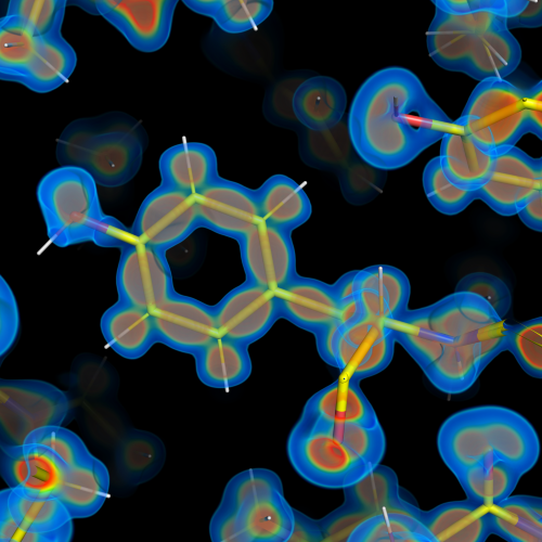
Refinement of the archaeal rubredoxin structure with the DiSCaMB transferable aspherical atom model (TAAM) library connected to the [refinement] software BUSTER directly reveals quantum mechanical phenomena. The image shows a valence electron deformation map with the rubredoxin protein model (sticks) surrounded by the electron density map (blue to red: low to high density). The map reveals electrons at the midpoint of chemical bonds and (valence) electron lone pairs.
© Ashwin Chari & Gleb Bourenkov
High-Brightness Data Collection
Achieving this resolution record required using one of the world's brightest X-ray radiation sources: the PETRA III storage ring at the Deutsches Elektronen-Synchrotron (DESY) in Hamburg, Germany. Data were collected at the European Molecular Biology Laboratory (EMBL)-operated P14 beamline.
Data collection and processing were driven in real time by Global Phasing’s automated workflow software. By calculating optimal multi-axis orientations, the software dynamically tailored a strategy that ensured all diffraction reflections were measured with high completeness and uniform multiplicity while actively steering the goniometer to prevent hardware shadows from blocking high-angle diffraction on the detector.
Minimizing Sample Damage
However, obtaining data at this level of resolution remains a major challenge, as intense X-ray irradiation inherently causes radiation damage that destroys the delicate structural details researchers aim to observe. They overcame this bottleneck by combining three crucial experimental advances during data collection:
-
Large Crystals: Using remarkably large crystals—600 × 500 × 250 µm³, compared to the typical 10–50 µm standard—increases the sample volume by three to five orders of magnitude. Because diffraction intensities scale with crystal volume, an equivalently lower radiation dose can be used. Moreover, a large crystal offers far more homogeneous diffraction properties than a collection of smaller ones.
-
Top-Hat Beam Profile: Instead of using a traditional Gaussian beam—which concentrates high intensity in the center and burns out the crystal unevenly—the beam shape was matched to the crystal size with a flat, "top-hat" intensity profile. This delivers uniform exposure across the entire crystal, preventing damage gradients so the entire sample degrades evenly at a much lower rate.
-
High-Efficiency Cadmium Telluride (CdTe) Detector: Using a DECTRIS EIGER2 X CdTe 16M detector equipped with CdTe sensors boosted quantum efficiency at high energies by more than 5 times compared to using Si sensors. This high efficiency, in combination with noise-free photon counting, allowed the team to collect data with minimal irradiation.
Together, these three strategies kept the total absorbed radiation dose down to just 0.5 MegaGray - 40 times lower than the conventional 20 MegaGray limit for protein crystallography - preserving the delicate sub-atomic details from being destroyed by radiation damage.
These findings have now been published in Acta Crystallographica Section D, Structural Biology.
Impact on Future Research
“Ultimately, this full experimental observation will enable the development of customized drugs for human diseases, as well as designer enzymes to catalyze chemical reactions,” says Dr. Ashwin Chari, head of the Structural Biochemistry and Mechanisms research group at the Max Planck Institute for Multidisciplinary Sciences in Göttingen (Germany). The long-term goal is to gain deep insights into enzyme-catalysed reactions to accelerate future progress in chemistry, materials science, and medicine.
References
Paknia, E., Flensburg, C., Chodkiewicz, M. L., Fogh, R. H., Keller, P., Vonrhein, C., Schulze-Briese, C., Dominiak, P. M., Bourenkov, G., Bricogne, G. & Chari, A. (2026): Towards routine accurate electron-density studies of biological macromolecules. Acta Crystallogr. D Struct. Biol. 82, 1044-1055, https://doi.org/10.1107/S2059798326007448
Every electron counts: most detailed protein structure to date revealed. Press Release, Max Planck Institute (2026). https://www.mpinat.mpg.de/5314392/rn_2604
¹Rubredoxin: A small, iron-binding protein found in bacteria and archaea that facilitates biological electron transfer. Because of its compact size and exceptional stability, crystallographers frequently use rubredoxin as a benchmark model system to test advanced X-ray techniques.
²Ångström (Å): A unit of length equal to 0.1 nanometer (nm) or 100 picometers (pm)—roughly the scale of a single atom—used by the scientific community to measure sub-atomic structures and chemical bond lengths.